一名八十歲女性病患,本身患有糖尿病,心血管疾病與老年癡呆症,因突發大量咖啡色嘔吐物伴隨上腹壓痛與坐立不安至急診就診,臨床理學檢查懷疑腹膜炎,電腦斷層攝影檢查發現腹內空氣與腹水。因懷疑中空臟器破裂,經過與家屬討論(消化性潰瘍穿孔須清洗腹膜,修補破洞; 減小手術傷害使用減孔腹腔鏡手術,若情況危急可能轉為開腹手術)
術中發現一巨大潰瘍位於十二指腸第一與第二部分交界處,穿孔處已波及超越十二指腸腔1/2圓周,且潰瘍遠端鄰近乳突部與總膽管,無法使用自動吻合器關閉,是故先行胃竇切除與Billroth-II 胃空腸吻合,十二指腸穿孔以v-lok進行閉合,並經腹壁放入導尿管權充十二指腸減壓造廔。
另外為減少術後全靜脈營養使用,以免體液過量造成組織水腫進而導致呼吸衰竭與吻合口腸壁腫脹使吻合處癒合不良,亦有做灌食空腸造廔 (通過傷口保護套直接目視進行,影片中未拍攝到)
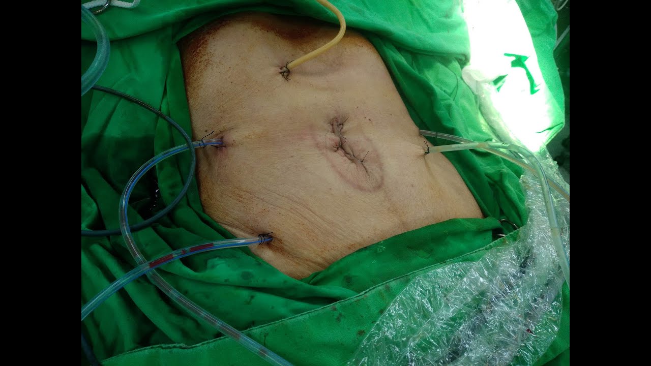
腹部兩側的5mm穿刺針傷口被用來放置引流管,為使十二指腸潰瘍修補處有良好引流,於右下腹另放置一條引流管。
術後患者順利拔管,轉至一般病房進行術後照護。
A 80-year-old female with previous history of Alzheimer’s disease, diabetes and CAD with regular dipyridamole use, brought to our emergency department due to massive coffee ground vomitus accompanied with restlessness and epigastric severe pain with tenderness.
CT showed massive free air with fluid collection at subhepatic and pelvis, after discussion the patient receive hybrid SILS peritoneal exploration for her peritonitis.
During operation massive kissing ulcer at the junction of duodenal bulb and 2nd portion was noted, with nearly transection of duodenum, the distal margin of ulcer is adjacent to CBD and pancreatic head, the gastroduodenal artery was obliterated within the periulcer necrotic tissue. Division of antrum was performed to fully expose the ulcer margin. The Billroth-II anastomosis was done laparoscopic given continuity of GI tract has to be maintained and a second-look operation to do re-anastomosis may be impractical given patient has no sign of hemodynamic instability or profound shock during anesthesia.
After attempts failed to lengthen the distal duodenal stump to fit the closure of autosuture, the ulcer was carefully evaluated. Even if instant conversion into laparotomy and Kocher maneuver was carried out, the ulcer may still had inadequate length to be sealed by auto suture device due to nearby common bile duct and ampulla. Therefore closure of distal duodenal stump with 1-0 v-lok braided suture and foley as decompressive duodenostomy for biliary diversion, aimed to facilitate healing.
To facilitate early feeding while avoid unnecessary IV fluid from TPN which may lead to interstitial edema, causing respiratory failure and/or compromising anastomosis, a feeding jejunostomy was made under direct vision with the wound retractor portion of the NELIS glove port still in place.
A total of 3000cc of lactated ringer was irrigated to cleanse the peritoneal cavity, 2 close drainage tube was inserted via the 5mm trocar aperture at bilateral side of abdominal wall. Additional close drainage at RLQ was inserted to ensure adequate drainage efficiency of the huge distal duodenum stump.
After the operation the anesthesiologist extubate the patient smoothly and the patient was transfer to ordinary ward for post-operative care.


コメント